Roles played by depressing synapses in neural circuits
Filed under:
Computational neuroscience
Joanna Jędrzejewska-Szmek (University of Warsaw Faculty of Physics), Jarosław Żygierewicz (University of Warsaw Faculty of Physics)
Synapses exhibiting short-term synaptic depression can be found in different locations in neural circuits [1], e.g. pyramid-pyramid and interneuron-pyramid connections. Based on these findings, two Wilson-Cowan type models with depressing synapses were constructed, following formalism presented in [2]. First one comprised a depressing positive feedback, the second one comprised a depressing negative feedback (synapse is between the inhibitory and excitatory population). The models' parameters were provided
by [1,3]. Both models generated oscillatory signals as predicted by [2,3].
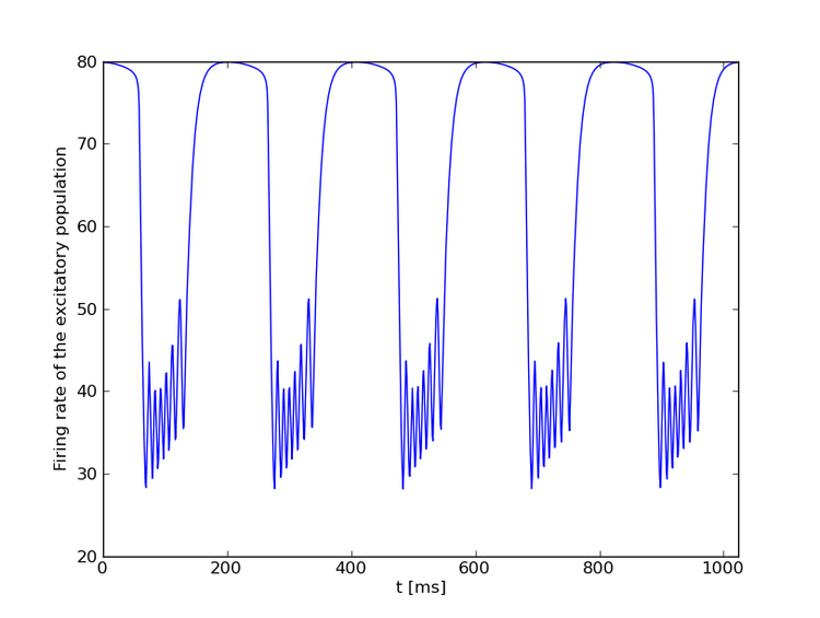
In case of both models a regime exists with high frequency oscillations (higher than 80 Hz) and high amplitude of oscillations corresponding to 'high-gamma' characteristics [4]. The model with depressing positive feedback showed three regimes: 1) oscillations in the theta band almost independent of external input of the excitatory population, 2) high-frequency oscillations with an envelope frequency in the theta band. In the third regime we observed oscillations in theta and 'high-gamma band'. An increase of the external input can abruptly change the oscillation frequency from the theta band to 'high-gamma'. Excitatory population firing rate is shown in the Figure below. One can see, that generated activity is similar to the theta/gamma oscillations [5] linked to short-term memory [5,6].
Short-term synaptic depression plays important role in generation of not only slow
oscillations activity [3] and theta waves, but also higher frequency phenomena such as 'high-gamma'. It
might also serve as a basis for coupling between the high- and low-frequency bands of
ongoing electrical activity in the human brain [5,6,7].
[1] Bannister AP, Thomson AM. Cerebral Cortex 2007
[2] Tsodyks MV, Pawelzik K, Markram H. Neural Computation 1998
[3] Melamed O, Barak O, Silberberg G, Markram H, Tsodyks M. Journal of Computational Neuroscience 2008
[4] Ray S, Maunsell JHR. PloS Biology 2011
[5] Lisman J. Hippocampus 2005
[6] Lisman J, Idiart MA. Science 1995
[7] Canolty RT, Edwards E, Dalal SS, Soltani M, Nagarajan SS, Kirsch HE, Berger MS, Barbaro NM, Knight RT. Science 2006

Preferred presentation format:
Poster
Topic:
Computational neuroscience

 Latest news for Neuroinformatics 2011
 Follow INCF on Twitter
